
Tiered Mentoring Program
Tracing ultra-structural neural connection through the retina
Dr. Jordan Renna
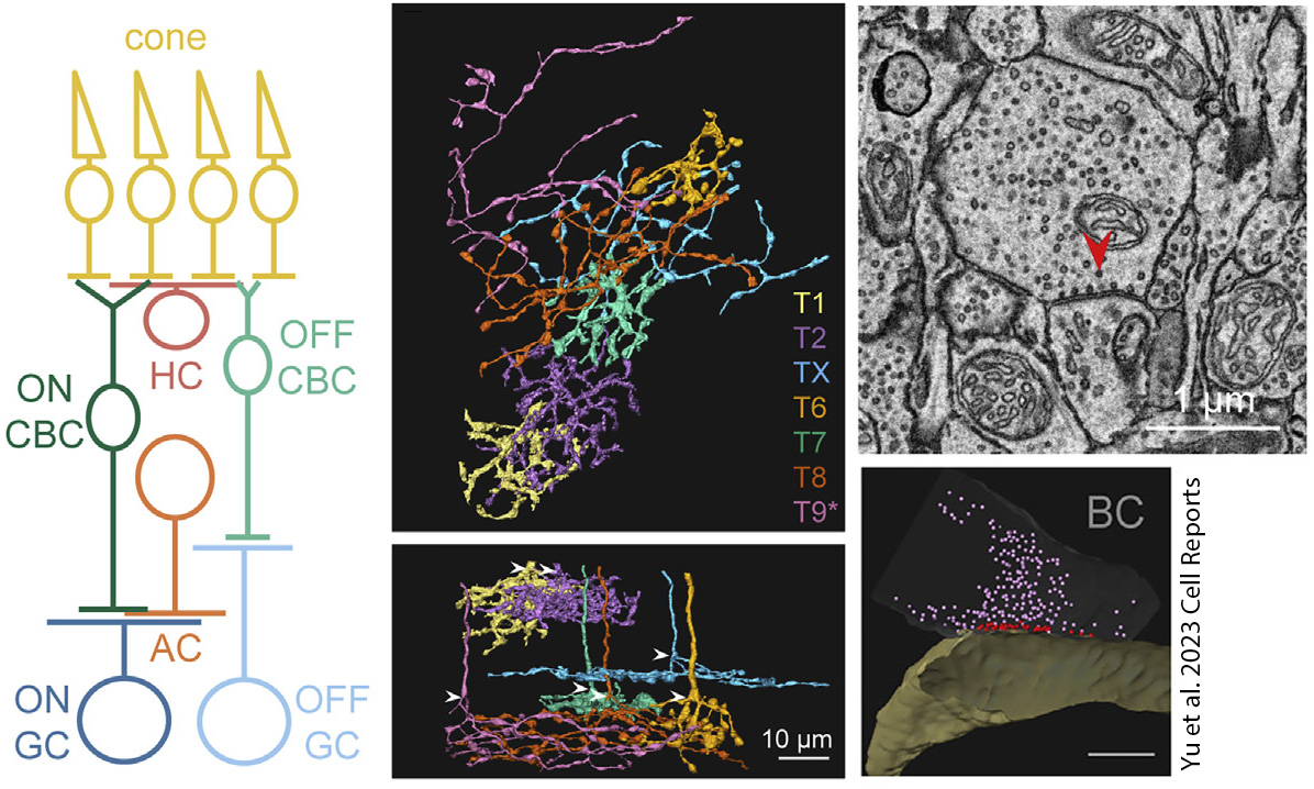 The Renna lab studies the neural circuits of the visual system as a window into how our brain processes information and guides human behavior. The retina is a thin layer of tissue in the back of the eye that detects light, converts it to an electrochemical signal that is filter through layers of complex connections before being sent to brain. My lab is interested in understand the ultra-structural intricacies of these retinal connections, and is using a large data set of serial block-face electron microscopy (SBEM) images to trace neurons and identify synaptic connections.
SBEM represents a cutting-edge approach that allows researchers to delve into the intricate details of this vital ocular structure. SBEM offers an unprecedented level of precision in visualizing the retina's complex architecture at nanometer-scale resolution. By sequentially removing thin sections of tissue and capturing high-resolution electron micrographs, scientists can reconstruct three-dimensional models of retinal layers, cell types, and synaptic connections. This technique not only provides insights into the structural intricacies of the retina but also aids in understanding how diseases like retinal degeneration or diabetic retinopathy affect its microanatomy.
This computer-based research project will involve using free online software to trace circuits and identify neuroanatomical structures through on online-database or SBEM images. It can be done remotely, or while on campus in the Renna lab. Students will also attend lab meetings and learn more about the retina and how our visual system functions.