
Discovering a little-understood cell in mice that opens a world of color
Dr. Jordan Renna is part of a team of scientists that has discovered that color vision in mice is far more complex than originally thought. Their findings could aid in developing treatments for human vision disorders and diseases.
The study was featured on the Jan. 3, 2018, cover of the prestigious journal Neuron.
Renna, an assistant professor of biology here, is on a team of scientists, led by Dr. Maureen Stabio, that discovered a new property of a little-understood cell in the eye called the M5. Stabio is an assistant professor in the Department of Cell and Developmental Biology at the University of Colorado School of Medicine.
The researchers knew that the M5 cell expressed a light-sensitive protein called melanopsin that allows mice to detect blue light. But as they investigated the function of the M5 cell, they discovered that the cell also compares signals from UV and green detecting opsins and then sends these integrated color signals to the brain for interpretation.
Function of retinal neurons
Renna and Stabio’s work focuses primarily on understanding the form and function of retinal neurons, including a group called intrinsically photosensitive retinal ganglion cells or ipRGCs that express the melanopsin protein, which includes the M5.

Dr. Renna Jordan
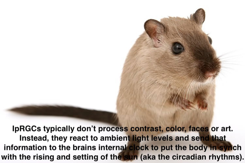
IpRGCs are involved in a kind of vision known as non-image forming vision because they typically don’t process contrast, color, faces, or art. Instead, they react to ambient light levels and send that information to the brain’s internal clock to put the body in synch with the rising and setting of the sun, also known as the circadian rhythms. The M5 cell was the least understood of the ipRGC types, until now.
Stabio, Renna and their colleagues found that the M5 cell might play a role in both image-forming and non-image-forming vision.
Surprising discovery
“You typically don’t think about color-coding functions in ipRGCs, since they are ambient light detectors,” Renna explains. “The biggest surprise of this work is that we found the M5 cell also can process color information. Mice are nocturnal and generally have poor vision. They navigate chiefly by using their nose and whiskers. What exactly they are doing with this color information remains to be discovered.
“We now know there is a class of neurons in the eye dedicated to blending image forming and non-image forming pigments and circuits used for different physiological and behavioral functions,” Renna continues. “This opens the door for researchers to begin to explore and potentially develop mechanisms to exploit this population of neurons for visual restoration in some disease models.”
Media contact: Lisa Craig, 330-972-7429 or lmc91@uakron.edu.